Unit 5
Protective Structure of the Central Nervous System:
A. Vertebrae (wait til lecture)
B. Meninges (p376-377)
1. Dura Mater
Epidural Space
2. Arachnoid Mater
Subarachnoid Space
3. Pia Mater
subdural hematoma (p376):
meningitis (p377):
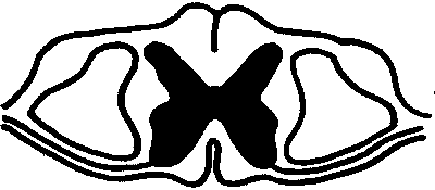
The Spinal Cord (p377-379):
A. Location (wait til lecture)
B. Structure of the spinal cord
1. Spinal Nerves (wait til lecture):
a. Mixed Nerves
b. Posterior Root
c. Anterior Root
2. Enlargements of Spinal Cord (see additional handout)
3. Type of tissue found in spinal cord:
a. White Matter
i. Grooves of Spinal Cord (p378):
Anterior Median Fissure:
Posterior Median Sulcus:
ii. Columns (funiculi):
Anterior Column:
Lateral Column:
Posterior Column:

b. Gray Matter (wait til lecture):
i. Horns (p378):
Anterior Horn
Lateral Horn
Posterior Horn

C. Function of Spinal Cord (p378):
Ascending tracts:
Descending tracts:
D. Reflexes (p383 - 385):
1. Pathway (Arc) Reflex:
2. Diagram of Reflex Arc:

The Brain (p387):
A. Four Major Parts:
1: Cerebrum
2: Diencephalon:
3: Brainstem (medulla oblongata, pons, and midbrain):
4: Cerebellum:
B. Cerebrum (p 387-397)
1. Structure
a. cerebral hemispheres- two large similar halves to the cerebrum.
b. corpus callosum- bridge of nerve fibers connecting cerebral hemispheres
Anencephaly (p388)
2. Surface of Cerebrum (p389)
a. convolutions (gyri or ridges):
Lissencephaly (p389)
b. Sulcus (pl. sulci):
c. fissures:
i. longitudinal fissure
ii. central sulcus
iii. lateral fissure
d. Lobes of Cerebrum (4 in each side)
i. frontal- anterior part of each hemisphre
ii. parietal- posterior to frontal lobe
iii. temporal- below lateral fissure/frontal lobe
iv. occipital- posterior part of cerebrum/above cerebellum
v. insula- covered by frontal, parietal, and temporal lobes
4. Function of cerebrum (p390):
a.
b.
c.
d.
5. Cerebral Cortex
- thin layer of gray batter covering the cerebrum
- brain power
- unmyelinated nerves covering white myelinated fibers
Functional regions of Cerebral Cortex (Cerebrum)
a. Primary Motor Areas
b. Primary Sensory Areas (somesthetic)
c. Associative Areas (problem solving, memory, speech etc...)
PRIMARY MOTOR AREAS: on frontal lobe along the central sulcus
(sends voluntary impulses to all of the body's skeletal muscle). (p 390; figure 11.16)
Apraxia:
**MOTOR AND SENSORY FIBERS CROSS OVER BEFORE GOING DOWN TO SPINAL CORD**
PRIMARY SENSORY AREAS (p392): interpret impulses from various receptors.
1. Parietal lobe- primary somesthetic cortex
2. Temporal lobe- auditory cortex
3. Occipital lobe- visual cortex Associative Areas (p392): cortical areas adjacent to primary sensory areas involved in recognition, memory, reasoning, verbalization, and emotional feelings. Found in every lobe. If damaged other areas can take over some duties.
Frontal lobes- (specifically: prefontal area)
parietal lobes-
temporal lobes-
occipital lobes-
6. other items
3 types of memory (p393)- ability to retain information
a) sensory memory- very short-term retention of sensory input while something is scanned, evaluated, and acted upon. Last less then one second. Caused by transient changes in membrane potentials.
b) SHORT-TERM MEMORY- information held for a few minutes. This memory is usually limited to 7 bits of information. (ex: telephone #) Caused by short-term changes in membrane potentials.
c) LONG-TERM MEMORY- Repetition of the information and the association of the new information from short-term to long-term memory. This information is stored for recall at any time. (ex remembering for a particular reason like a test etc...)
Cross-referenced in cortex
1. declarative-
2. procedural-
SLEEP- cerebral cortex inactive
RIGHT AND LEFT CORTEX
RIGHT HEMISPHERE
*spatial perception
*recognition of faces
*musical ability
LEFT HEMISPHERE
*mathematics
*speech
concussion (p395)
Cerebral Palsy (p395)
Stroke (p395)
7. Ventricles (p394-397):
Lateral ventricles:
Third ventricle:
Fourth Ventricle:
Cerebrospinal Fluid:
**read about the function of basal ganglia (p394) and the limbic system (p401)
**Review Table 11.7 (p405)
C. DIENCHEPHALON (p398-401)
*surrounds third ventricle
*the part of the brain between the brainstem and the cerebrum
*covered by the cerebral hemispheres
1. Main Parts of the Diencephalon (p398-400)
a. Thalamus- forms sides of third ventricle
b. Hypothalamus- forms floor of third ventricle (p400)
i.
ii.
iii.
c. Pituitary Gland- inferior part of the diencephalon on the stalk called infundibulum: size of a pea
D. BRAINSTEM (p401-403)
1. Parts of brainstem
a. medulla Oblongata (p402)
b. Pons (p402)
c. Midbrain (mesencephalon):- inferior to diecephalon
RETICULAR FORMATION
*group of nuclei distributed throughout the brainstem
*receives sensory information from the face
*these neurons play an active role in arousing and maintaining consciousness
*it forms the sleep/wake cycle
E. CEREBELLUM (p403-405)
1. Location:
2. Structure:
3. Function:
Peduncles- 3 paired bundles of nerve tract connecting cerebellum to the medulla and pons
Damage to Cerebellum:
4. BRAIN WAVES (p406)
EEG (electroenephalogram) measures electrical activity of cerebral cortex. Most of the time there is no particular pattern, at other times specific patterns can be detected.
Alpha waves: (8-13 cps) awake, eyes closed, resting
Beta: (13 or higher cps) awake, eyes open, intense mental activity
Theta: (4-8 cps) usually in children or stress, frustration and possible brain disorders in adults.
Delta: (4 or less cps) deep sleep
No waves: DEAD
EEG Diagnose- epilepsy, trauma, disease, tumors and bruises


